


(1) Overview
Introduction
Trabecular bone is a region of bone with a spongy, honeycomb-like structure. Analysis of trabecular bone provides important information to accurately characterize bone tissue, helping in researching bone diseases and investigating the progress of bone formation. Bone density, another bone characteristic, reveals the amount of mineral per volume of bone, but does not provide information directly about bone morphology. In contrast, trabecular thickness (Tb.Th) and trabecular spacing (Tb.Sp), two important parameters of trabecular structure, which are the average thickness of bone and none-bone, respectively, give more value information about bone structure to assess the effectiveness of bone forming agents [, , , , ]. Previous studies used diverse methods to estimate trabecular thickness, resulting in different, potentially non-reliable results. Standardization of trabecular thickness measurement is of paramount importance, consequently, trabecular thickness should be computed by a well-defined procedure.
The oldest method to calculate the average thickness of thin structures is histomorphometry. Thereby the thickness is calculated indirectly as a half of the area divided by its perimeter with an assumption based on the two-dimensional model []. This method can be incorrect, for example, when obtained points and intersections for the area and measurements of the perimeter are not representative for the object. Cruz-Orive proposed a surface-based analysis of trabecular thickness with a model assumption, in which the trabecular thickness at any point is measured by the distance from that point to the opposite surface []. However, this surface-based definition tends to induce inaccuracy problems for arbitrary and non-ideal structures. To assess the thickness of any two-dimensional object of any shape, Garrahan et al. introduced a solution to estimate the local thickness by fitting the maximal circles to every point inside the structure []. A similar method for three-dimensional structure analysis using the definition of the volume-based thickness was developed by Hildebrand and Rüegsegger and applied in the well-known open source software BoneJ [, , ]. In that way, the local thickness at one point is computed as the diameter of the greatest sphere that fits within the structure and contains the point. This algorithm accepts a binary mask input. Accordingly, the Euclidean distance of each point in the foreground to the closest background one is measured and shown as intensity at each point in the distance map [] (Figure 1). From that, the local thickness map of the structure is produced, of which intensity at a point is the value of the local thickness at the respective point in the object. In turn, the average trabecular thickness is defined as arithmetic mean value of the local thicknesses at all points in the structure. The algorithm based on the definition of Hildebrand and Rüegsegger shows good performance for extraction of the quantitative values with high resolution and informative inputs such as micro-computed tomography (µCT). In comparison with other software such as CT Analyser and Scanco, BoneJ provides similar results of trabecular thickness measurements with variations of up to 20% []. A reason for these differences could occur due to different approximations required by the algorithms to derive the results in acceptable computation times. Therefore, the need for a simple, well-defined and accurate reference algorithm without approximations to calculate trabecular thickness and spacing is apparent.
Example of a trabecular bone with a scale bar of 2mm and the respective measurements. a) Trabecular bone b) Binary mask c) Distance map d) Local thickness map.
In this work, we present a database of µCT bone images of a sheep femur as well as a simple reference brute force implementation for accurate trabecular thickness and spacing computations, which is derived from the volume-based thickness concept of Hildebrand and Rüegsegger. The algorithm is designed to be suitable for massively parallel computing because voxel values can be computed independently from each other. The accuracy of the reference implementation without approximations is validated with artificial datasets of ellipsoids. Furthermore, we assess how the correctness of trabecular thickness measurements depends on the voxel size and show that sub-voxel processing or upsampling processing can be used to increase the accuracy at the expense of computation time. The reference algorithm is applied to the introduced µCT database covering a wide range of trabecular thickness and spacing. The results are correlated with those from the current de-facto reference implementation, BoneJ, over a large distribution of trabecular thickness and spacing, showing strong agreement. However, the computation of the brute force method has time complexity O(n6) for cubic volumes with the size of each dimension of n and is therefore prohibitive for high resolution µCT datasets. We encourage scientists to develop fast parallel graphic processing unit accelerated (GPU-accelerated) algorithms to compute trabecular bone thickness and spacing, either accurately or approximately with well-understood error bounds. The accuracy of these methods can be assessed by using the provided datasets and the reference brute force implementation. The intermediate data from the brute force method are also provided to enable verification of their intermediate steps.
Implementation and Architecture
µCT datasets
40 datasets were cropped from a µCT scan of a femur of a 60-month-old female sheep, which was from a study about an animal model for aortic aneurysms []. The scan is 1,600 × 1,600 × 800 equilateral voxels, which was acquired by a µCT (MILabs B. V., Utrecht, The Netherlands) with a voltage of 55 kV and a current of 0.17 mA with 1,440 projections. Three-dimensional (3D) bone images were cut at varying positions of the µCT scan by using the software Imalytics Preclinical (Gremse-IT GmbH, Aachen, Germany) to get different rectangular forms, which have distinct trabecular thickness and spacing (Figure 2).
Examples of bone images with different forms and trabecular thickness. 3D rendering (upper panel) and 2D axial slice view (lower panel), respectively.
Our introduced database of bone images consists of 40 three-dimensional bone images in the nifti file format (.nii) with the image size of 100 × 100 × 100 voxels, voxel size of 20 × 20 × 20 µm3. Along with each bone image, the intermediate results, which are computed by both the brute force method and BoneJ, are also available.
Bone Segmentations
Since BoneJ and the brute force method require a binary mask as input, the bone images were segmented by using a single thresholding segmentation. The thresholding value was set as 2,000 HU (Hounsfield Units) and applied on the HU-calibrated µCT scans. The resulting binary mask includes trabecular bone as the foreground and the none-bone part as the background and is also attached in the dataset as stated in the section µCT dataset.
Brute force algorithm
To analyse the effect of voxelization, particularly for thin structures, we upsampled the input data using trilinear interpolation by a factor of two, meaning the voxels size is reduced by two. This upsampling was performed before applying the threshold to generate the binary mask. The thickness and spacing computations were performed with and without upsampling.
Moreover, the original bone images were downsampled with a factor of 0.5 and then upsampled with a factor of two. The thicknesses of the downsampled bone images and the downsampled-upsampled bone images are computed with the brute force method and compared together with the thicknesses of original images to examine the enhancement of accuracy.
Thickness measurements
The original volume-based trabecular thickness definition can be expressed by the formula 1 and Figure 3. According to the definition, the distance map value of a voxel describes the radius of the largest sphere contained in the foreground and centered at the voxel. From the distance map, the local thickness map is computed which describes the diameter of the largest sphere contained in the foreground including the voxel. The local thickness at point  is equal to twice the highest intensity in the distance map, point
  , that is bigger than the Euclidean distance between those two points as the following equations 2 and 3:
A surface of the object Ω. Measurement of volume-based thickness at the point inside a surface of the object Ω.


where , are points inside the object Ω, r is the radius of the sphere sph (, r) with the center at ,  is a point outside the object, DM () is the distance map at the point , LTM() is the local thickness at the point , Ω ϵ R3 is the set of all points in the object,
  is the Euclidean distance between and ,
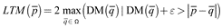
  is the Euclidean distance between and , and ɛ is a defined error, an important factor to account for border voxels, which are close to the surfaces of spheres. The value ɛ is selected experimentally as 0.5, a good value to include accurately border voxels without requiring a clean-up step as in BoneJ.
The brute force implementation runs over all other voxels in parallel to search for the maximum containing sphere, according to the equation 3. Therefore, it provides well-defined results without approximations at the expense of computation time and can be used as a reference algorithm.
Implementation
The algorithm was implemented in C++ with the standard library and Open Multi-Processing statements. The code contains 64 lines of code for the local thickness map calculation, 66 lines of code for distance map calculation with five primary functions and 1,900 lines of code for helper functions (Figure 4). Main functions are:
Workflows of the brute force algorithm.
CreateMask() creates a binary mask of the input image.
DistanceMapBruteForce() calculates the distance map
LocalDistanceMapBruteForceSearch() searches for the biggest sphere that contains a give voxel
LocalThicknessMapBruteForce() calculates the local thickness map
Average() calculates the mean value of the local thickness map
Generation of artificial datasets
For validation of the brute force algorithm, we assumed a fixed ellipsoid with diameters 100 × 100 × 300 mm3. A set of 500 3D binary images are created by generating voxelized binary masks of 500 images of an ideal ellipsoid in a cube, defined by an ellipsoid formula, sampling the fixed ellipsoid. The binary datasets are composed of 50 subsets differing in voxel sizes, in which 10 images of each subset share the same voxel size, but different relative positions of the ellipsoid in the cube. The whole of datasets varies namely in grid sizes of Z dimension from 15 to 309 voxels with increments between subsets of six voxels, leading to decreasing voxel sizes, from 40 mm to 1.942 mm. The binary images are inputted to the brute force method and BoneJ to produce the local thickness map of each ellipsoid cube image. The mean thickness of a subset of ellipsoid cube images with a certain voxel size is correspondingly evaluated and considered as the mean thickness of the ellipsoid with that voxel size with averaged error in trabecular thickness of the ellipsoid due to voxelization. Then, these results are compared to the thickness of the respective ideal ellipsoid of the same voxel size, as an exact value, which is computed by the closed formula by Hildebrand and Rüegsegger.
Quality control
The algorithm was tested with 500 ellipsoid cube images covering a wide range of voxel sizes from 1.942 mm to 40 mm. Furthermore, the code was also tested with the whole provided database of 40 scanned bone images with a broad range of trabecular thickness and spacing.
Artificial ellipsoid cube datasets
To validate the brute force algorithm, we used artificially generated images with different voxel sizes of a fixed ellipsoid with diameters 100 × 100 × 300 mm3. The trabecular thickness inside the respective ideal ellipsoid is computed with a closed formula of Hildebrand and Rüegsegger and compared with the results from the brute force algorithm and BoneJ. Finally, the trabecular thickness and spacing of the 40 bone samples were calculated using brute force and BoneJ and correlated to examine their accuracies, supporting that the brute force can be used to validate the future methods for trabecular thickness measurements.
Since the local thickness map computation involves voxelization, a discretization error in the range of the voxel size is expected in both the distance map and the local thickness map. In Figure 5 the error in the mean thickness of ellipsoids appears systematic due to the voxelization. Generally, voxelization may result in a large relative error if large voxel sizes relative to the structure sizes are used. Figures 5b and 5c show that the discretization error becomes worse for bigger voxel sizes. Figure 5c illustrates that the brute force method and BoneJ involve large relative errors up to 20% for big voxel sizes. Both the brute force algorithm and BoneJ require voxel sizes under 5.4 mm and 4.4 mm or 111 voxels and 135 voxels in the Z dimension, respectively, to reach relative errors below 4%. The correctness of the brute force and BoneJ improves for the smaller voxel sizes, suggesting the usage of the upsampling technique with these methods for accuracy enhancement in future applications.
Accuracy of thickness computations on ellipsoid. a) Slice through the analytically derived local thickness map of the ellipsoid with diameters 100 × 100 × 300 mm3. The red color has a higher intensity, corresponding to a bigger local thickness. b) Mean local thickness as a function of voxel size in comparison with the exact values, which were mathematically calculated per voxel. Coarse voxels cause an underestimation of the thickness. c) The relative error to the exact value increases with the voxel size. BoneJ and the brute force algorithm have similar relative errors which increase with the growth of the voxel size.
Scanned bone datasets
Trabecular thickness and spacing were computed for the bone images using the brute-force method and BoneJ. Furthermore, the intermediate and final results were analyzed for original and upsampled datasets. With the same binary mask input, the brute force method and BoneJ produced identical distance maps but the local thickness maps differed slightly, possibly owing to a tradeoff in BoneJ favoring computation time over measurement accuracy. The 40 bone images showed a broad distribution of values for trabecular thickness (236 µm to 1,469 µm) and spacing (326 µm to 3,790 µm) as shown in Figure 6, emphasizing that the provided dataset is well-suited to examine the accuracies of trabecular thickness and spacing computations. Figure 6a shows a strong correlation for the trabecular thickness of the original brute force method and BoneJ as well as for the upsampled brute force method and BoneJ with correlation coefficients of 0.98 and 0.99, respectively. The Bland-Altman plots in Figure 6c suggest that the results of the brute force method are always slightly higher than those of BoneJ, by a difference on the order of the voxel size. Their difference tends to increase for thicker structures, is in the range of the voxel size and can be attributed to the compromise in BoneJ between the computation time and the thickness correctness. The trends are shared for the upsampled methods in Figure 6e with smaller mean difference 11.1 µm. For trabecular spacing, the mean difference is higher without an apparent systematic trend (Figure 6d, 6f).
Comparison between brute force and BoneJ for 40 bone datasets. Strong correlation between brute force and BoneJ appears for trabecular thickness (a) and trabecular spacing (b), with and without upsampling. Bland-Altman plots for trabecular thickness (c) and trabecular spacing (d) show systematically lower values for BoneJ compared to brute force, on the order of the voxel size. Bland-Altman plots for upsampled results (e, f) show stronger agreement between BoneJ and brute force.
To confirm the effect of voxelization, the thicknesses of the downsampled bone images with the voxel size of 40 µm and the downsampled-upsampled bone images with the voxel size of 20 µm are compared to those of the original images. Figure 7 demonstrates that the relative deviations of the thicknesses of the downsampled images from the thicknesses of original images are mostly bigger than those of the downsampled-upsampled images. In other words, with upsampling, the thicknesses of the downsampled-upsampled images are closer to those of the original images, which is in strong agreement with Figure 5c. The upsampling enhances certainly the accuracy of thickness measurement.
Relative deviation of thicknesses of downsampled bone images and downsampled-upsampled bone images from those of the original images by the brute force method.
(2) Availability
Operating system
The code may be compiled on any platform and C++ compiler but was only tested with Windows 7 and 10 with Visual Studio 2019.
Programming language
C++
Additional system requirements
CMmake Version 3.1 or newer is required to generate the project files, for example, for Visual Studio 2019, which can then be used to compile the code. Our code only uses C++ with the standard library and OpenMP statements.
Dependencies
N/A
List of contributors
N/A
Software location
Archive
Name: Figshare
Persistent identifier
For software: https://doi.org/10.6084/m9.figshare.17209319
For bone dataset: https://doi.org/10.6084/m9.figshare.9864677
Licence: MIT licence
Publisher: Thi-Ngoc-Thu Nguyen, Felix Gremse
Version published: 3.0
Date published: 15/09/21
Code repository
Name: Code Ocean
Identifier: https://doi.org/10.24433/CO.3957929.v3
Licence: MIT licence
Date published: 15/09/21
Language
English
(3) Reuse potential
We implemented a simple brute force algorithm of 130 lines of code with the helper functions of 1,900 lines of code to compute trabecular thickness and spacing according to the concept of Hildebrand and Rüegsegger. Its accuracy was evaluated by the comparison of the outputs with those of BoneJ on artificial ellipsoid datasets of different resolution and on bone datasets. BoneJ with a certain approximation and a complicated algorithm of more than 1,500 lines of code along with the helper functions of thousands of lines of code and the straightforward brute force method without approximations produce very similar results. The both methods show systematic underestimation of trabecular thickness which increases with the voxel sizes. This phenomenon can be attributed to the similar binarization steps inside brute force and BoneJ. By upsampling the ellipsoid images using trilinear interpolation, utilizing information on the subvoxel level, binary images are generated at higher resolution, resulting in more accurate values for trabecular thickness, indicating that upsampling can reduce the systematic underestimation caused by the binarization. This is also completely in agreement with the experiment of downsampled bone images and downsampled-upsampled bone images.
Using 40 cubic datasets of different bone regions of a sheep femur with a broad range of trabecular thickness and spacing, we show that brute force and BoneJ strongly correlate in their estimations of trabecular thickness and spacing. Their results indicate differences on the order of the voxel size which can be attributed to implementation details. Upsampling reduces the mean difference between the two algorithms, supporting the idea that upsampling can increase the accuracy by performing the binarization on a finer grid. This is illustrated with the probability p-value < 0.001 in Supplementary File S1 against the null hypothesis of mean difference between two methods. The paired two-sample t-test demonstrates that the mean of the differences between thicknesses of bone images by the upsampled brute force method and BoneJ is smaller than those by the original brute force method and BoneJ. The thickness measurements by the upsampled methods reach closer to the exact values.
Unfortunately, upsampling strongly increases the computation time to generate trabecular thickness and spacing. Particularly, the brute force method with the time complexity O(n6) becomes extremely time-consuming for the large input images. Faster methods could be implemented using GPU-acceleration or by performing a hill-climbing search to find the largest enclosing sphere for each voxel. A pyramid approach could be used to avoid local maxima during hill-climbing. While we intend to provide a simple reference algorithm without approximation for such performance optimizations, our analysis also shows that BoneJ with a certain approximation performs well for both artificial and real datasets, delivering values close to our brute force algorithm, supporting its wide-spread use.
Researchers are encouraged to download the databases for reuse from Figshare. Dataset usage is meant for evaluation of the accuracy of methods to compute trabecular thickness and spacing. The trabecular thickness results of future methods can be compared with those of the brute force method over the whole dataset to get the distribution of their difference over a range of data. The intermediate data from the dataset such as binary masks, distance maps and local thickness maps of the brute force method can also be used to verify intermediate steps of the future methods. Moreover, the researchers can test the brute force algorithm with images beyond the provided database.
Additional File
The additional file for this article can be found as follows:
Supplementary File S1Paired two-sample t-test for comparison of the means of the differences of thicknesses of the bone images calculated by the original brute force method and BoneJ and those by the upsampled brute force method and BoneJ. P(T<=t) two-tail is the important probability p-Value. DOI: https://doi.org/10.5334/jors.360.s1